Trump threatens steeper India tariffs over Russian oil

Donald Trump says he will significantly increase tariffs on goods from India through Russian oil purchases, while protecting the new Delhi interests and calling the US President as “unfair”.
Trump said that after announcing that Washington will apply 25 percent tariff to the goods imported from the country that started last Friday last Friday, he negotiated with India.
India encountered pressure from the West, including the United States, to get away from Moscow after invading Ukraine in the early 2022 of Russia. Yeni Delhi resisted this pressure with Russia showing its long -standing ties and economic needs.
“India is not only buying a large amount of Russian oil, but they sell it in the open market for large snow for most of the purchased oil.
“Therefore, I will greatly increase the tariff paid by India to the USA.”
He didn’t explain what the tariff would be.
At the weekend, two Indian government sources told Reuters to continue to buy oil from Russia despite Trump’s threats. Sources did not want to be defined because of the sensitivity of the issue.
Washington showed geopolitical disagreements with India to explain why it could not reach a trade agreement with the new Delhi.
Apart from India’s ties with Russia, Trump has published the BRICS group of developing nations, which is an important part of India.
The Indian Foreign Ministry spokesman said India’s “will take all necessary measures to protect national interests and economic security”.
“In this background, the target of India is unfair and unreasonable,” he said.
India, India, India, India began to import oil from Russia, because the Ukrainian conflict was directed to Europe after the outbreak, he said.
The Sözcü said that India’s imports were to provide affordable energy costs for Indian consumers, and that it was a necessity with a global market status ”.
In a statement, the West, especially the European Union’s bilateral trade with Russia, said: “The nations criticizing India themselves fell into trade with Russia.”
India was also disappointed by Trump receiving credit many times to an Indian-Pakistan ceasefire announced on social media on May 10th. The ceasefire stopped the days of hostility between nuclear armed Asian neighbors.
India’s position is that the new Delhi and Islamabad should solve the problems without direct participation.
Trump made a trade agreement with Pakistan.
Elsewhere, Switzerland said that he was ready to make a “more attractive offer in his trade talks with Washington, in a statement on Monday, after a crisis meeting aiming to prevent a tariff on Swiss goods that threaten to influence the export -guided economy.
Federal Council, the country’s administrative cabin, said that beyond the last date of August 7, it is determined that President Trump was determined to have negotiations with the US beyond the last date of 7 August, which was determined to enter into force.
“Switzerland is ready to offer a more attractive offer to this new stage, taking into account our concerns and trying to alleviate the current tariff status,” he said.
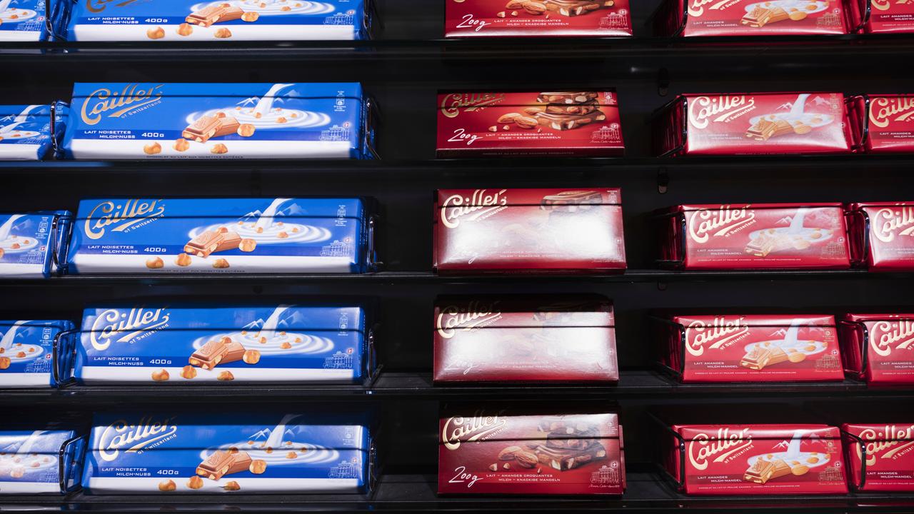
Switzerland counts the US as the best export market for medicine, watches, machines and chocolates.
Australian Associated Press is a beating heart of Australian news. AAP has been the only independent national Newswire of Australia and has been providing reliable and fast news content to the media industry, the government and the corporate sector for 85 years. We inform Australia.


